
Fig. 1.
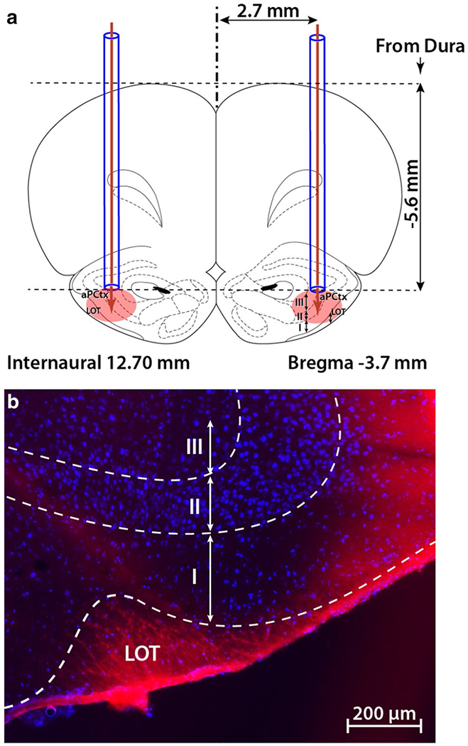
Site of intracranial injection in the anterior piriform cortex (aPCtx). a Two cannula guides were symmetrically implanted (coordinates: AP – 3.7 mm from Bregma, ML ± 2.7 mm and DV – 5.6 mm from dura), and 1 μl of drug or control vehicle (ACSF) was bilaterally injected (glucose, insulin, or MgTx). The coronal drawing was adapted from the Paxinos and Watson atlas (2007), plate 7. b A representative example of one brain section through the aPCtx, 30 min following the injection of 1 μl Evans blue. LOT lateral olfactory tract; I, II, III layers of the aPCtx; blue DAPI nuclear stain