FIGURE 2.
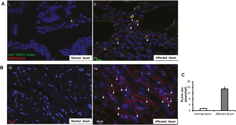
Complex formation between latent TGF-β1 and GRP78 is increased in SEMF of fibrostenotic ileum in vivo compared with normal ileum in the same patient. A, Co-immunostaining of GRP78 and latent TGF-β1 in affected ileum compared with histologically normal ileum (i and ii). B, Direct protein-protein interaction between latent TGF-β1 and GRP78 is increased in affected ileum compared with normal ileum in the same patient with fibrostenotic Crohn’s disease. Proximity ligation hybridization assay was used to demonstrate direct protein interaction within 15 nm as indicated by the red chromogen. Images are representative of the normal and fibrostenotic ileum in 5 different patients. C, Data are expressed as pixels per high power field in each of 10 consecutive high power fields with similar numbers of cells. *Denotes P < 0.05 between normal ileum and affected ileum. Results are expressed as mean ± SEM of 5 patients.

