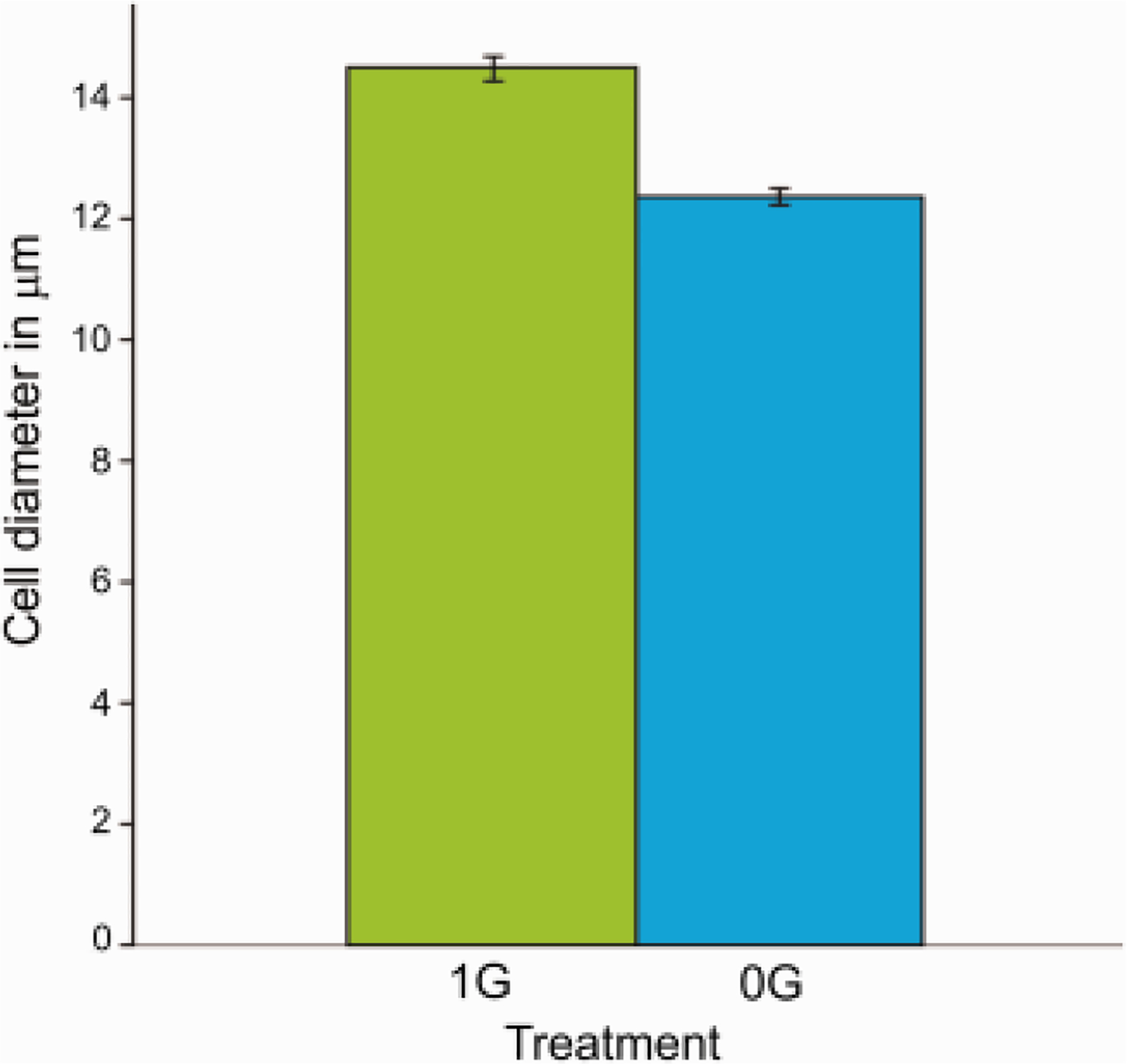
Fig. 3. The cell diameter of OLs derived from Hu embryonic brain is reduced after exposure to sim-μG.

Cells cultured in OSM for 4 days revealed significantly smaller cell body diameter when cultured under sim-μG conditions as compared to cells cultured under 1G conditions. This phenomenon might be attributed to cytoskeleton and cell adhesion changes. Values are expressed as mean + SEM of two independent experiments. Data were analyzed using unpaired t-test **: p<0.01 ***: p<0.001 vs. respective control.
