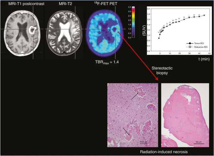
Fig. 1.
A 70-year-old patient with an anaplastic astrocytoma. Contrast-enhanced MRI 31 months after radiation therapy suggests tumor progression. In contrast, O-(2-[18F]-fluoroethyl)-l-tyrosine (FET) PET shows only slight metabolic activity, and the time–activity curve shows a constantly increasing FET uptake, consistent with treatment-related changes. After a stereotactic biopsy, histological examination yielded signs of radiation-induced necrosis (hematoxylin and eosin staining, original magnification ×200; scale bar, 50 mm). Brain parenchyma shows reactive changes and blood vessels with thickened hyalinized walls (arrows; hematoxylin and eosin staining, original magnification ×100; scale bar, 1000 mm; reproduced from Galldiks et al.,59 with permission from Oxford University Press).

