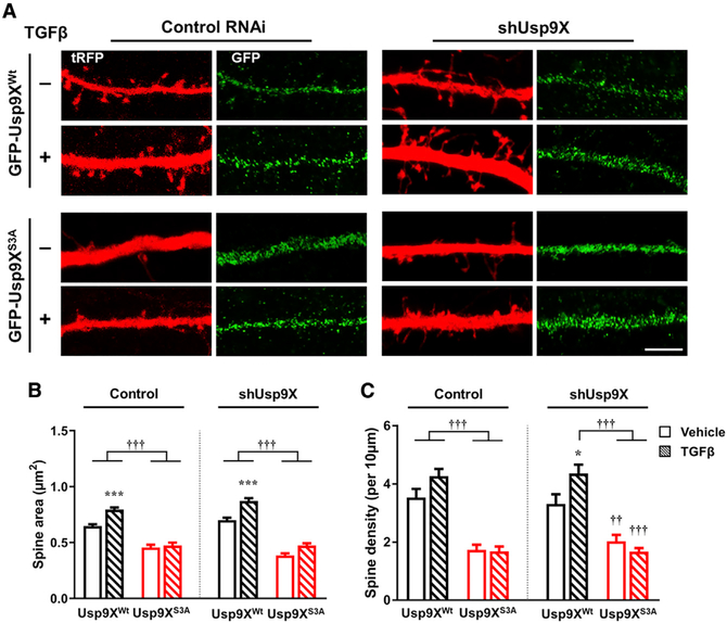
Figure 6. Replacement of RNAi-Resistant GFP-Usp9XWt, but Not GFP-Usp9XS3A, Induces TGF-β-Dependent Spine Enlargement.
(A) Confocal images of neurons transfected with control RNAi (control) or shUsp9X and GFP-Usp9XWt or GFP-Usp9XS3A (green) and subsequently treated with vehicle or TGF-β (20 ng/mL) for 1 h in a dendritic region outlined by tRFP (red) expression. Scale bar, 5 μm.
(B and C) Spine head area (B) and density (C) in control or shUsp9X and GFP-Usp9XWt (black bars) or GFP-Usp9XS3A (red bars) with vehicle (plain pattern) or TGF-β (comb pattern) (n = 12–18 neurons for each group). Vehicle versus TGF-β: *p < 0.05, ***p < 0.001; GFP-Usp9XWt versus GFP-Usp9XS3A: ††p < 0.01, †††p < 0.001 by one-way ANOVA followed by non-parametric statistical analysis. TGF-β (−), vehicle; TGF-β (+), 20 ng/mL TGF-β for 1 h. All data are presented as mean ± SEM.

