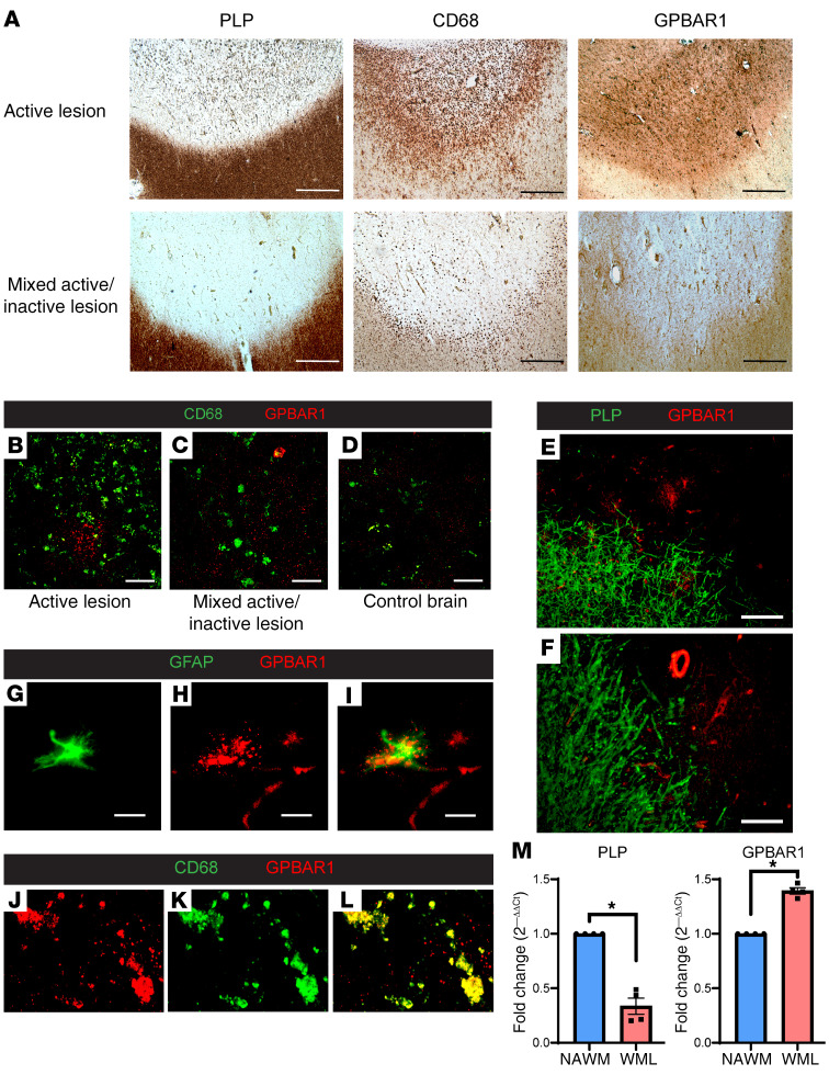
Figure 4. The cell-surface bile acid receptor GPBAR1 is detected in demyelinating lesions in PMS brains.
(A) Immunohistochemistry for PLP and CD68 identified an active and a mixed active/inactive MS WML, with GPBAR1+ cells detected within both of these lesions. Comparison of GPBAR1 staining (red) in an active lesion (B), a mixed active/inactive lesion (C), and control brain (D) revealed increased staining in MS lesions compared with control white matter. Double immunostaining for PLP (green) and GPBAR1 (red) shows the presence of GPBAR1+ cells (E) and vessels (F) in areas of demyelination. (G–I) Double immunostaining using GFAP (green) and GPBAR1 (red) demonstrates GPBAR1 staining in GFAP+ astrocytes in MS lesions. (J–L) Double immunostaining with CD68 (green) and GPBAR1 (red) demonstrates GPBAR1 staining in CD68+ macrophages in MS lesions. (M) Comparison of PLP and GPBAR1 gene expression in MS WML and NAWM revealed decreased PLP expression within the lesions with increased expression of GPBAR1, similar to findings noted on immunohistochemistry (n = 4 in each group). Scale bars: 200 μm (A); 100 μm (B–F); 20 μm (G–I). *P < 0.05, by 2-tailed Mann-Whitney U test. Data in M represent the mean ± SEM.

