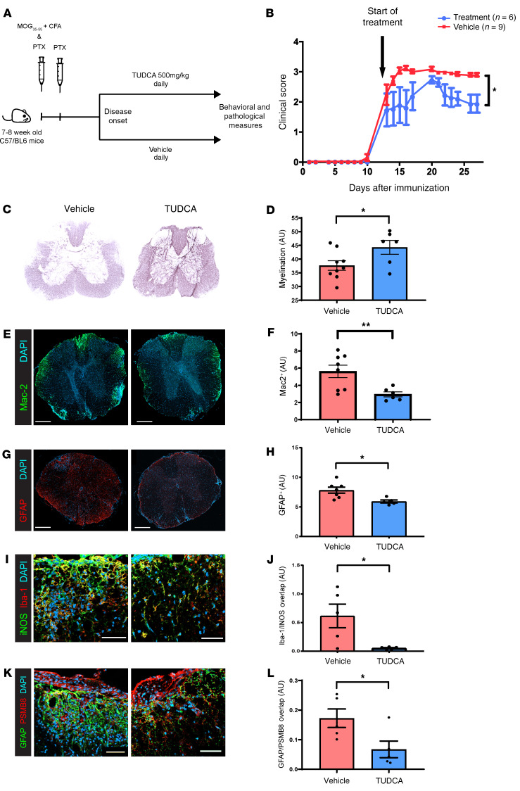
Figure 6. TUDCA supplementation ameliorates EAE.
(A) Eight- to 9-week-old female C57/BL6 mice were subcutaneously immunized with MOG35–55 and CFA in addition to intraperitoneal injection of pertussis toxin (PTX) on day 0 and day 2. Mice were then monitored for signs of EAE and, at disease onset, were randomized to oral gavage with either TUDCA or vehicle until day 28 after immunization. (B) Behavioral scores demonstrate that TUDCA treatment resulted in reduced severity of EAE disease. Data were derived from a representative experiment of 1 of 3 independent experiments and represent the mean ± SEM. *P < 0.05, by Mann-Whitney U test. (C and D) Representative images of Black-gold staining of spinal cords of mice with EAE treated with either TUDCA or vehicle show reduced demyelination with TUDCA treatment. Quantification is shown in the plot in D. (E and F) Immunostaining for infiltrating myeloid cells (Mac-2+) demonstrated reduced infiltration in the TUDCA-treated group compared with the control. Quantification is shown in the plot in F. (G and H) Staining for GFAP showed reduced astrocytosis in the TUDCA-treated group compared with the control. This is quantified in the plot in H. (I and J) Staining for Iba-1 and iNOS shows a decrease in INOS+Iba-1+ cells (M1 macrophages/microglia) in the TUDCA-treated group compared with the control. Quantification is shown in the plot in J. (K and L) Images show a reduced number of PSMβ8+GFAP+ cells (neurotoxic A1 astrocytes) in the TUDCA-treated group compared with the control. This is quantified in the plot in L. Scale bars: 200 μm (E and G); 50 μm (I and K). *P < 0.05 and **P < 0.005, by unpaired, 2-tailed Student’s t test (D, F, H, J, and L). Data represent the mean ± SEM (D, F, H, J, and L).

