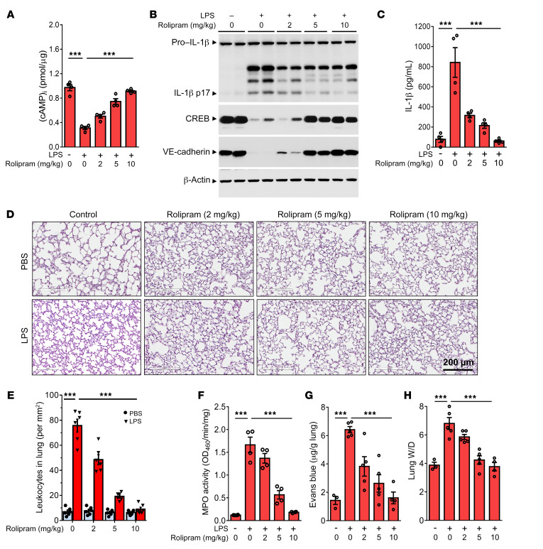
Figure 4. Augmentation of cAMP signaling by rolipram suppresses inflammatory lung injury.
(A) C57BL/6J mice (n = 4) were administrated with rolipram (2, 5, and 10 mg/kg) for 1 hour, and injected intraperitoneally with LPS (12 mg/kg) for 1 day. Intracellular cAMP levels of the lung lysates were measured by ELISA. (B) Lung lysates were prepared for expression detection of IL-1β maturation, CREB, and VE-cadherin by Western blot. (C) Measurement of IL-1β levels in mouse serum, as determined by ELISA (n = 4). (D) H&E staining of the lung is shown (scale bar: 200 μm; n = 3). (E) Quantitative analysis for leukocyte infiltration in lungs (n = 6). (F) Quantitative analysis of neutrophil infiltration by measurement of lung tissue MPO activity (n = 4). (G) Lung vascular permeability is detected by EBD leakage in the lungs (n = 3–5). Extracted dye contents in the formamide extracts are quantified by measuring at 620 nm. (H) The ratios of the wet lung to dry lung weight were assessed (n = 3–5). Results are shown as mean ± SEM. ***P < 0.001. Statistics obtained from ANOVA.

