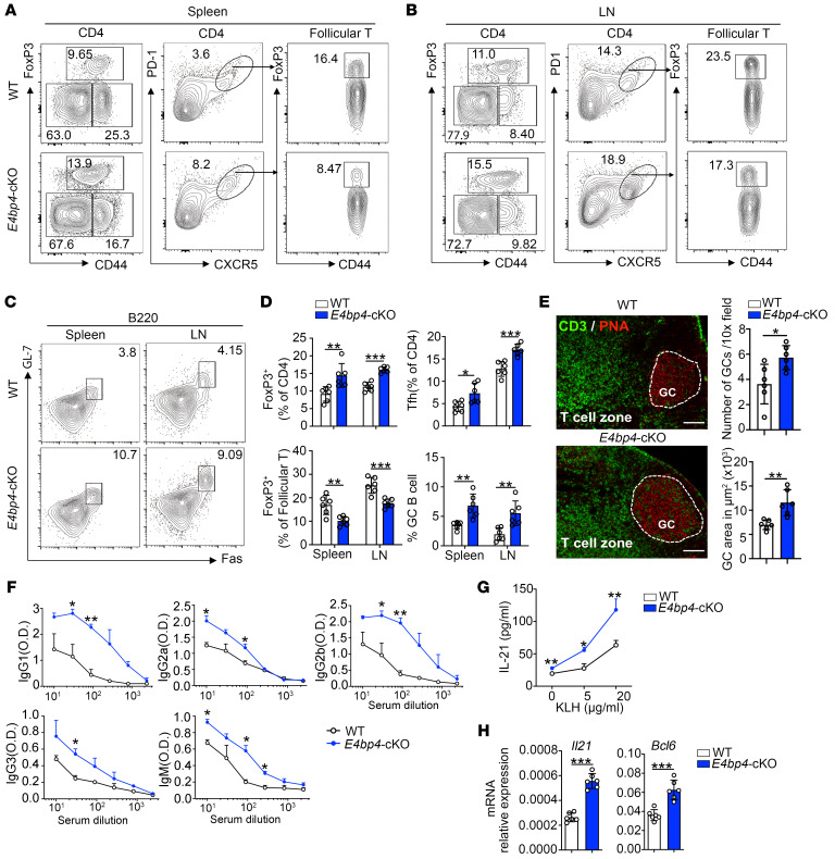
Figure 4. E4BP4 deficiency promotes antigen-specific GC responses.
Age-matched WT or E4bp4-cKO mice were immunized with KLH for 14 days, then lymph node and spleen cells were harvested. (A and B) Flow cytometric analysis of FoxP3+ Treg cells, CD4+CXCR5+PD-1+ Tfh cells, and FoxP3+ Tfr cells in follicular T cells. (C) Analysis of B220+Fas+GL-7+ GC B cells. (D) Summary of the percentage of FoxP3+ Treg cells, Tfh cells, and FoxP3+ Tfr cells in follicular T cells, as described in A and B, and GC B cells, as described in C. (E) Immunofluorescence of GCs from WT and E4bp4-cKO mice, representative images of CD3 and PNA staining of LNs. Scale bars: 200 μm. Quantification of PNA+ GC areas and number of PNA+ follicles per lymph node. (F) Detection of serum anti-KLH–specific IgG1, IgG2a, IgG2b, IgG3, and IgM by ELISA. (G) Draining lymph node cells were restimulated with KLH for 3 days, and supernatant IL-21 expression was detected by ELISA. (H) Il21 and Bcl6 mRNA expression levels were analyzed by qPCR (n = 6). Data are representative of 4 independent experiments. Student’s t test. *P < 0.05; **P < 0.01; ***P < 0.001.

