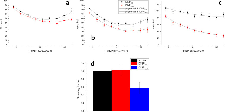
Figure 4.
Cytotoxicity of IONP on HeLa cells. (a–c) Proliferation kinetics of HeLa cells incubated with IONP during 48, 72 and 96 h. One-way ANOVA statistical analysis revealed a significant difference between treated groups and control; Two-way ANOVA statistical analysis proved significant difference between IONPs and DOX-IONPs (P < 0.0001 for 48 h; P < 0.0001 for 72 h; P < 0.0001 for 96 h). Also, the presence of DOX in the construct induced a significant reduction of proliferation, compared to equivalent concentrations of IONPCO (P = 0.0003 for 48 h; P < 0.0001 for 72 h; P < 0.0001 for 96 h). (d) Clonogenic survival of HeLa cells seeded in the colony formation assay after exposure to 100 μg/mL IONP for 16 h. Data are presented as percentage of untreated control and are shown as mean ± SEM (n = 3).

