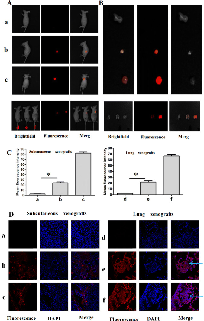
Fig. 4.
In vivo identification of miR-155-5p and fluorescence imaging of cancer cells in xenografts models. a Imaging the subcutaneous and lung xenografts after injection of CS-MB via the tail veins. Subcutaneous xenografts model, a: A549 treated with CS-RS MB, b:A549 treated with CS-miR-155-5p MB, c: H446 treated with CS-miR-155-5p MB. Lung xenografts model, d: A549 treated with CS-RS MB, e:A549 treated with CS-miR-155-5p MB, f: H446 treated with CS-miR-155-5p MB. Groups a and d were used as the negative controls. b Imaging the xenografts after removal. c Fluorescence intensity was analyzed after injection (n = 8) (*p < 0.05). d Confocal microscopy imaging of the xenografts tissues after transfection with CS-miR155-5p MB or CS-RS MB (red). Cell nuclei were stained by DAPI (blue). Scale bar 25, 50 μm. Arrow: planting nodule and cancer cells

