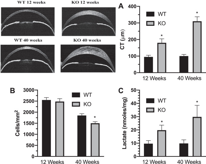
Figure 6.
Corneal [lactate] is increased in a mouse model of congenital hereditary endothelial dystrophy. (A) OCT images of corneal cross-sections illustrating differences in CT plotted for 12- and 40-week-old wild-type and Slc4a11 knock out mice (n = 9 mice per condition). (B) Endothelial cell density (n = 3 mice per condition). (C) Corneal [lactate], n = 3 mice per condition. *P < 0.05, significantly from wild-type mice.

