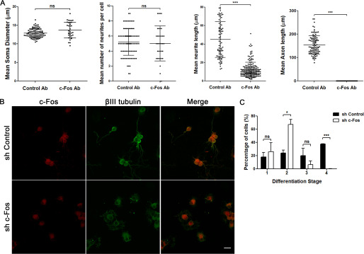
Figure 2.
Blocking the activity or the expression of c-Fos impairs neuronal differentiation. A, cells were profected with anti c-Fos (c-Fos Ab) or with a nonrelated anti-mouse IgG antibody as a control (Control Ab) 2 h after seeding using BioPORTER and then were fixed after 48 h of culture. The different morphological aspects were quantified from microscopy images, using ImageJ software, and are shown as the mean ± S.D. (error bars) in each case. Student's t test statistical analysis was performed using GraphPad Software. ***, p < 0.001; n.s., nonsignificant; in each experiment, n = 40 cells from each condition were examined. Results of one of three independent experiments performed are shown. B, cells were infected at the initiation of the culture with lentiviral particles designed to express an anti c-Fos shRNA or an shRNA with a scrambled sequence of c-Fos as a control. After 48 h of culture, cells were fixed and immunostained with an anti-c-Fos antibody (red, first column) and an anti βIII-tubulin antibody (green, second column). The third column shows the merge between both labels. Scale bar, 20 μm. C, morphological quantification of neuronal differentiation stages in both c-Fos and scrambled infected cells was performed using ImageJ software. The graph shows the mean number of cells ± S.D. in each case. Student's t test statistical analysis was performed using GraphPad software. *, p < 0.05; ***, p < 0.001; n.s., nonsignificant; n = 198 cells for scrambled shRNA–infected cells, n = 254 cells for c-Fos shRNA-infected cells. Results of one of three independent experiments performed are shown.

