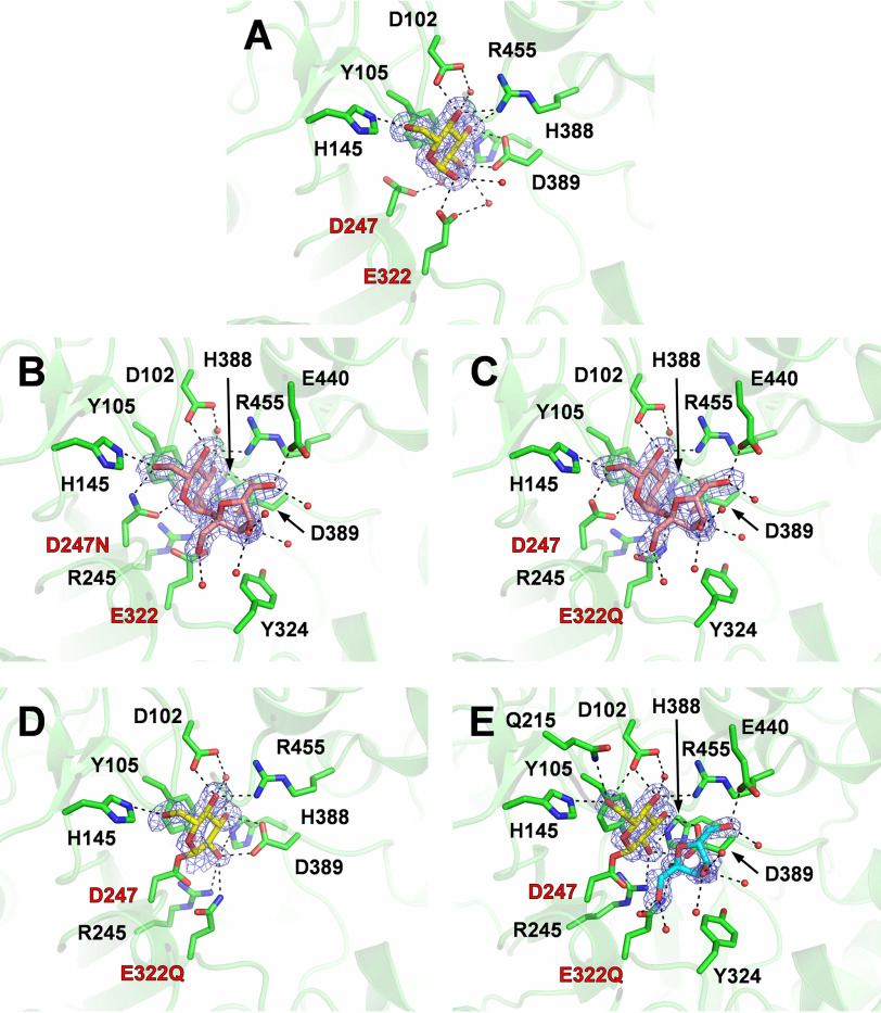
Figure 2.
Active sites of BmSUH complexes with substrates, intermediates, and products. Active-site structures of BmSUH-Glc (A), D247N-Suc (B), E322Q-Suc (C), E322Q-GlcF (D), and E322Q-GlcF-Fru (E). The side chains of the amino acid residues and ligands are indicated as stick models, and water molecules interacting with ligands are shown as red spheres. |Fo| − |Fc| omit maps (contoured at 2 σ) for ligands and hydrogen bonds are shown as blue mesh and a dashed line, respectively. Labels of catalytic residues are highlighted in red. Colors used are as follows: amino acid residues (green), glucose and its covalent intermediate (yellow), sucrose (pink), and fructose (cyan).

