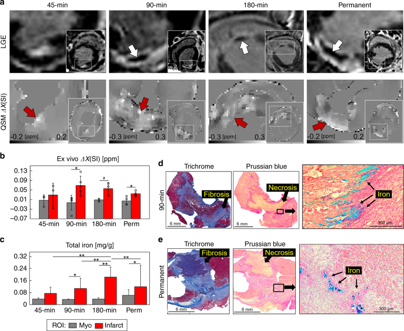
Fig. 1. Infarct magnetic susceptibility is associated with elevated tissue iron content.
At 1 week post-infarction (a), late gadolinium enhanced (LGE) MRI shows hyperintense signal in the infarct region corresponding to uptake of gadolinium contrast agent. In total, 90, 180 min, and permanent infarcts were larger with microvascular obstruction, as indicated by hypointense regions in the infarct core (white arrows). Images show views of the infarct region and left ventricular short axis (insets). Infarcts show a paramagnetic shift in ex vivo quantitative susceptibility maps (QSM) for each infarct group compared with remote (myo) regions. b There was a significant infarct paramagnetic shift in 90 min (P = 0.012) and permanent (P = 0.025) infarct groups. Magnetic susceptibility measurements in 45 min (n = 3), 90 min (n = 5), 180 min (n = 3), and permanent (n = 4) infarct groups were from independent whole heart ex vivo specimens, regions of interest (ROI) were compared using a two-tailed two-sample t test for each infarct group. c Infarct total iron concentration was significantly elevated in 90 min (P = 0.001), 180 min (P < 0.0001), and permanent (P = 0.015) infarcts compared with remote myocardium. In total, 45 min (n = 31, 24), 90 min (n = 32, 32), 180 min (n = 48, 37), and permanent (n = 30, 50) measurements were from independent tissue samples (myo, infarct), tissue regions and infarct groups were compared using two-way analysis of variance (ANOVA) with Tukey’s HSD post-hoc test. d Representative histological findings in a 90 min reperfused infarct. Trichrome stain shows a nontransmural infarct, fibrosis, and nonviable myocytes at the core of the infarct. Prussian blue staining shows iron accumulation (black arrows) at the transition zone between necrotic myocytes and mixed viable myocytes suggesting an active immune response originating outside the infarct core. e Representative histological findings in a permanent occlusion infarct. Trichrome stain shows fibrosis and transmural infarct. Prussian blue shows iron deposits at the peripheral infarct fibrotic regions. Representative histology from each infarct group and remote myocardium are shown in Supplementary Fig. 6. Histology was repeated independently on multiple tissue sections (n > 5) for each whole heart specimen (n = 15) showing similar histopathological findings for each infarct group. The results are reported as mean ± SD. #P = 0.094 and significance *P < 0.05 and **P < 0.001. Source data for b and c are provided as a Source data file.

