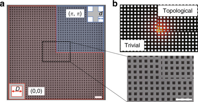
Fig. 1. Design of the topological nanocavity.

a Scanning electron microscopy image of a fabricated 2D topological PhC cavity in a square shape. The inset on the right shows an enlarged image around the corner. The scale bar is 1µm. The topological nanocavity consists of two topologically distinct PhCs, which are indicated by the red and blue areas. They have different unit cells, as shown in the insets. d and D are the lengths of the squares in the blue and red unit cells, in which D=2d. b Electric field profile of the topological corner state
