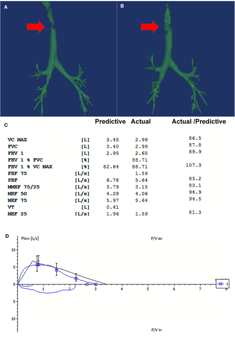
Figure 2.
The repeated three-dimensional computed tomography (3D-CT) of the trachea and the spirometry at 16-month follow-up. (A) There was partial absence at the right side of the trachea on the 3D-CT on admission (Red arrow pointed to the fistula). (B) The repeated CT at 16-month follow-up showed that the fistula was covered by the flap grafting (Red arrow). (C) The spirometry at 16-month follow-up showed that the ventilatory function was normal. (D) The flow-volume loop showed that no fixed or dynamic airway obstruction existed. Good patency and stableness of large airway was maintained after the treatment.

