Fig. 2.

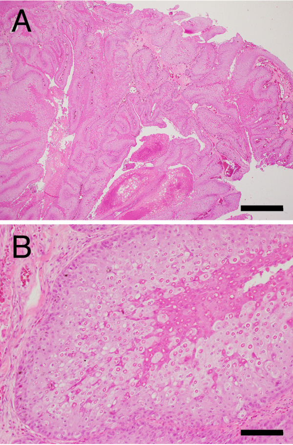
Histopathology of the cutaneous nodules of Case No. 1. hematoxylin and eosin staining. (A) Low-magnification. Bar=1 mm. (B) Higher magnification of (A). Bar=100 µm.

Histopathology of the cutaneous nodules of Case No. 1. hematoxylin and eosin staining. (A) Low-magnification. Bar=1 mm. (B) Higher magnification of (A). Bar=100 µm.