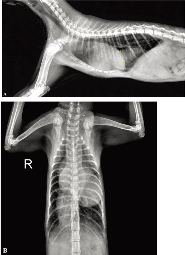
Fig. 1.

(A) Right lateral and (B) ventrodorsal radiograph of a seven-month-old Korean shorthair cat. Radiographs demonstrate severe cardiomegaly and pruning of the peripheral pulmonary artery with increased radiolucency of the lung field. A tubular soft-tissue density is visible on the caudoventral thorax extending from the heart to the diaphragm. To make a reasonable comparison before and after the medication treatment, the ratio of the diameter of the CVC to the length of 5th thoracic vertebral body (CVC:V) was measured. On this radiograph, the CVC:V ratio was 6.82. Dotted line, diameter of the CVC; solid line, length of 5th thoracic vertebral body.
