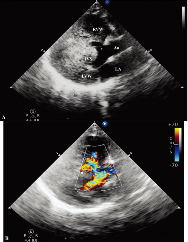
Fig. 2.

Right parasternal long axis, five-chamber view echocardiogram. (A) Two-dimensional echocardiography shows a 3-mm perimembranous ventricular septal defect. The right ventricular wall is markedly hypertrophied. (B) The color-Doppler demonstrates the turbulent flow across the left ventricular outflow tract and the right ventricle. Ao, aorta; LVW, left ventricular wall; RVW, right ventricular wall; IVS, interventricular septum; LA, left atrium. Asterisk (*), ventricular septal defect.
