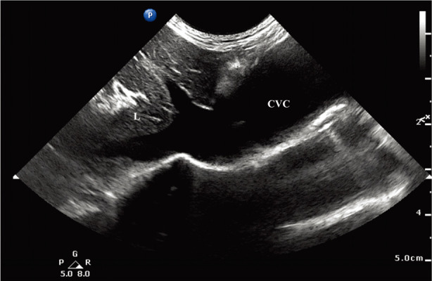
Fig. 4.

Ultrasound images obtained from right caudal thorax with the left-hand side of the image directed cranially. A 3 cm large tortuous vessel is seen on the caudal thorax that was identified as CVC since it is connected to the right atrium cranially and to the hepatic veins caudally. A small amount of pleural effusion is seen around the field. L, liver; CVC, caudal vena cava.
