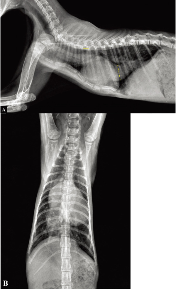
Fig. 5.

Right lateral (A) and ventrodorsal (B) thoracic radiographs following 1 month after starting medication. Reduction of the CVC was noted, as compared to a previous thoracic radiograph. On this radiograph, the CVC:V ratio was 4.09. Dotted line, diameter of the CVC; solid line, length of 5th thoracic vertebral body.
