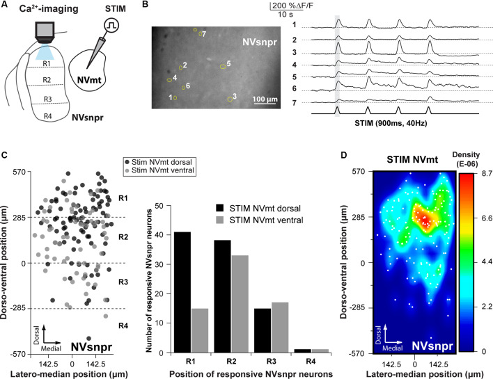
Figure 4.
Distribution of NVsnpr neurons activated by stimulation of NVmt. (A) Schematic drawing of the brainstem slice preparation and the experimental conditions used. (B) Photomicrograph showing the position of responsive cells in NVsnpr following electrical stimulation of dorsal NVmt and their synchronous Ca2+ responses. Gray vertical line indicates the length of the train of stimulation. The bottom trace shows a stimulus artifact. (C) Distribution of cells responding to stimulation of both NVmt-D and NVmt-V throughout NVsnpr (Left) and count of the number of cells per division in NVsnpr (Right). (D) Heatmap representing the density of responsive neurons in NVsnpr following stimulation of NVmt-D and NVmt-V pooled together. Abbreviations: Stim, stimulation; NVsnpr, trigeminal main sensory nucleus; NVmt, trigeminal motor nucleus.

