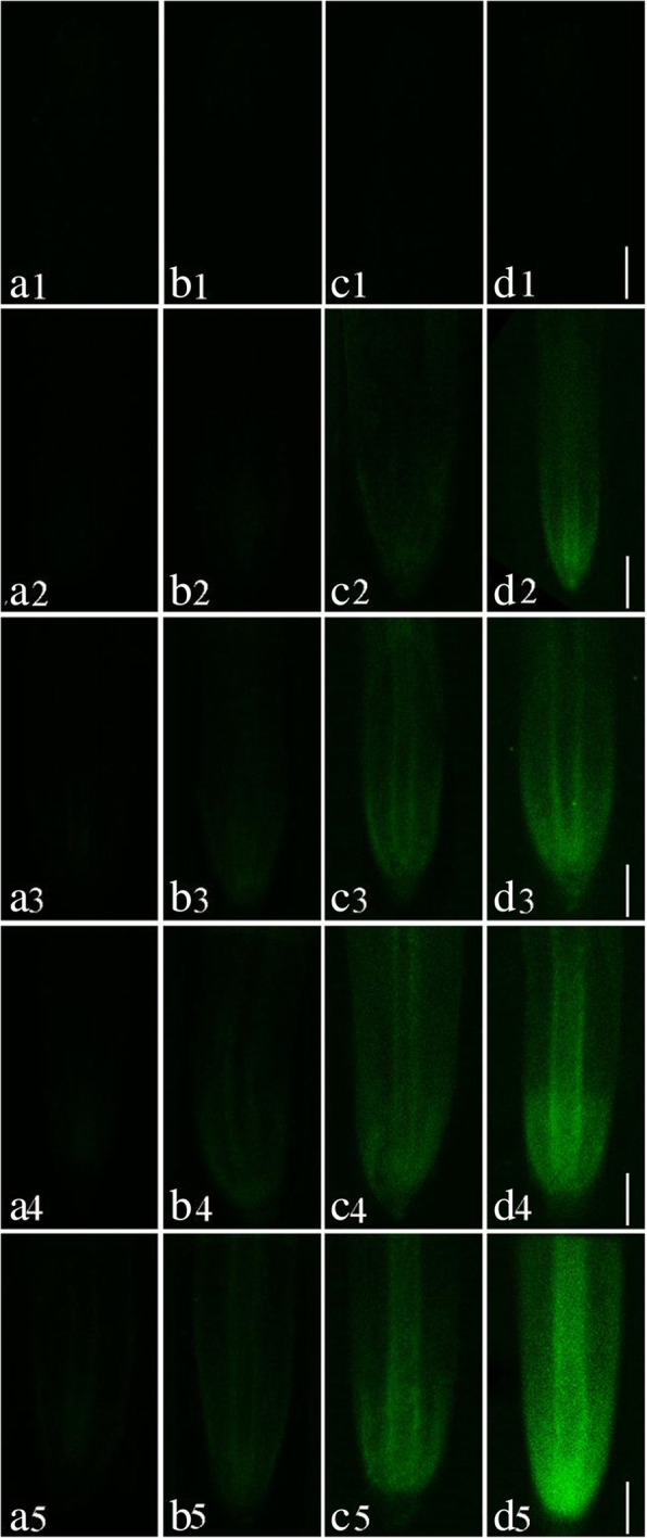
Fig. 7.

Micrographs of S. babylonica roots using Leadmium Green AM dye at longitudinal sections of roots exposed to different Pb concentrations (0, 1, 10, 50, or 100 μmol/L) for different treatment times (3, 6, 12, and 24 h). a1–d1: Control without Pb for 3, 6, 12, and 24 h; a2–d2: 1 μmol/L Pb for 3, 6, 12, and 24 h; a3–d3: 10 μmol/L Pb for 3, 6, 12, and 24 h; a4–d4: 50 μmol/L Pb for 3, 6, 12, and 24 h; a5–d5: 100 μmol/L Pb for 3, 6, 12, and 24 h. Scale bar = 1 mm
