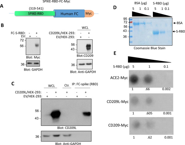
Figure 4: CD209L and CD209 bind to SARS-CoV-2-S-RBD.

(A) Schematic of Fc-CoV-2-S protein is shown. (B) Expression of Fc-CoV-2-S and CD209L in HEK-293 cells. (C) Immunoprecipitation assay demonstrates the binding of CD209L with Fc-CoV-2-S protein. (D) Coomassie blue stain of CoV-2-S-RBD is shown. (E) CoV-2-S-RBD applied onto PFVD membrane with varying concentrations via Dot blot apparatus. The membranes after blocking with 5%BSA were incubated with cell lysates derived from HEK-293 cells expressing ACE-2-Myc, CD209L or CD209 and binding of ACE2, CD209L and CD209 to CoV-2-S-RBD was detected anti-Myc antibody.
