
FIGURE 5.
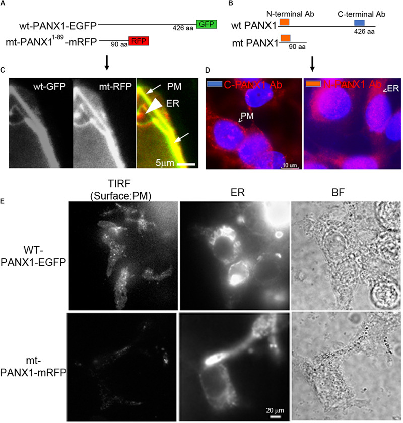
Localization of PANX1. (A,B) Schematic of fluorescent WT and mt PANX1 constructs (A) and the N and C-terminal epitopes recognized by anti-PANX1 antibodies (Ab) (B). (C) Localization of WT PANX1-EGFP and mt PANX1-mRFP in transfected HEK cells. (D) Localization of endogenous PANX1 in PC-3 cells using N- or C-terminal specific Abs. Nuclear DAPI stain is depicted as blue. (E) TIRF imaging on HEK cells transfected by WT PANX1-EGFP or mt PANX1-mRFP constructs. WT PANX1-EGFP localizes in the PM and the ER, while mt PANX1-mRFP only in the ER.