
FIGURE 9.
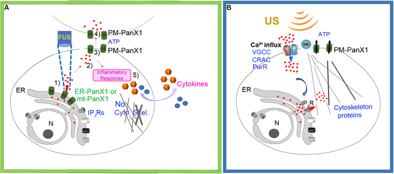
(A) Schematic of new working model FUS-dependent response mechanisms in PC-3 invasive cancer cells. (1) FUS stimulation activates ER localized mechanosensitive PANX1 resulting in internal Ca2+ release from ER stores. (2) This cytoplasmic Ca2+ signal stimulates ATP release through PANX1 PM channels. (3) The released ATP acts on purineregic receptors, many in adjacent cells. (4) This results in a propagating extracellular Ca2+ wave which spreads through the cell population possibly via PM PANX1 or opening of PM Ca2+ channels. (5) FUS stimulation also results in secretion of cytokines/chemokines. (B) Schematic of currently accepted working model based largely on conventional US stimulation with no proposed role for internal ER Ca2+ release but rather a link to US energy transduction to ER mediated by IP3R. MB, Microbubbles.