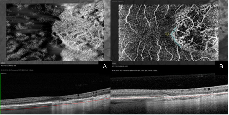
Fig. 3.
Choroidal changes in SC (continued). The hypoperfusion seen in Fig. 2 leads to a window defect responsible for the “White on Black effect” in the Haller’s Layer seen here at the top of (a). b displays the same defect at the level of the retinal pigment epithelium, showing that the area corresponding to the foveola was spared. The OCT angiograms are projections of the retina at an imaginary line running 140 μm inferiorly and parallel to the basal membrane in (a) and through the manually segmented retinal pigment epithelium in (b), shown in the corresponding OCT B-Scans below

