Figure 1.
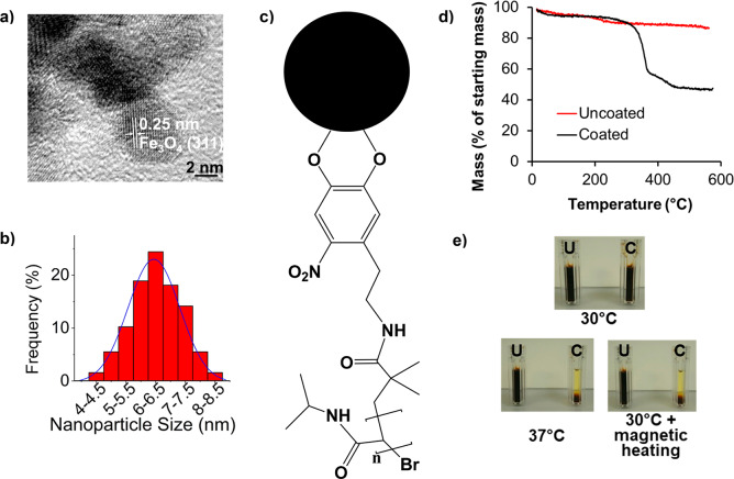
Characterization of polymer-coated SPIONs. (a) High-resolution transmission electron micrograph of nanoparticles and (b) the associated size distribution, as determined from lower-magnification micrographs. For calculation of the shown lattice plane distance, see also Figure S1. A Gaussian fit over the SPION size distribution is shown.19 (c) Schematic of nitrodopamine-terminated PNIPAM and its attachment to the SPION surface (the black sphere denotes a SPION). (d) TGA of 10 mg each of uncoated and PNIPAM-coated SPION samples, which were heated under nitrogen gas at a ramp rate of 10 °C/min between 0 and 600 °C. (e) Photographs of 10 mg/mL PNIPAM-coated (C) and uncoated (U) SPIONs incubated at the indicated temperatures for 30 min in plastic cuvettes. For magnetic heating, the sample was exposed to a 108 kHz/0.67 T alternating magnetic field before quick transfer to the cuvette for photographing.

