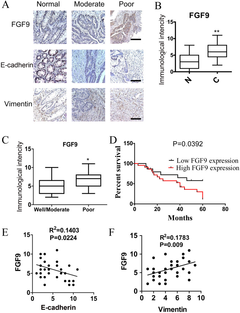
Fig. 4.
FGF9 predicts poor survival in patients with CRC. (A) Immunohistochemical staining of FGF9, E-cadherin and Vimentin in paraffin-embedded human CRC tissues. Scale bars: 25 μm. (B) Immunohistochemical scores for FGF9 in normal colorectal mucosa and CRC tissues (n=37). (C) Immunohistochemical staining of FGF9 in well-, moderately and poorly differentiated CRC tissues. (D) Kaplan–Meier survival curves of CRC patients with low (n=15) and high (n=22) FGF9 expression. FGF9 was classified as high expression if the immunostaining score is more than four, otherwise classified as low expression. (E) Correlation test of immunostaining intensity between FGF9 and E-cadherin. (F) Correlation test of immunostaining intensity between FGF9 and Vimentin. *P<0.05, **P<0.01.

