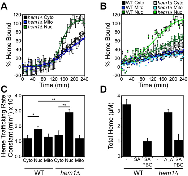
Fig. 3.

ALA synthase (Hem1) negatively regulates mitochondrial–nuclear heme trafficking. (A) hem1Δ cells expressing HS1 in the cytosol (black), nucleus (green), or mitochondria (blue) were pulsed with a bolus of ALA to initiate heme synthesis and the rates of heme trafficking to the indicated subcellular locations were monitored by measuring the fractional saturation of HS1 over time, as in Fig. 2. (B) WT and hem1Δ cells expressing the HS1 sensor were pre-cultured with SA to deplete heme and pulsed with a bolus of PBG to re-initiate heme synthesis. The rates of heme trafficking to the indicated subcellular locations were monitored by measuring the fractional saturation of HS1 over time, as in Fig. 2. Data shown in A and B are mean±s.d. of n=3 cultures (C) Heme trafficking rate constants for the indicated compartments of WT and hem1Δ strains, based on data represented in B. Data are mean±s.d. of triplicate cultures. *P<0.05, **P<0.001 (one-way ANOVA with Dunnett's post-hoc test). (D) Analysis of total heme in WT and hem1Δ strains treated with SA, ALA or PBG. Data represent the mean±s.d. of triplicate cultures.
