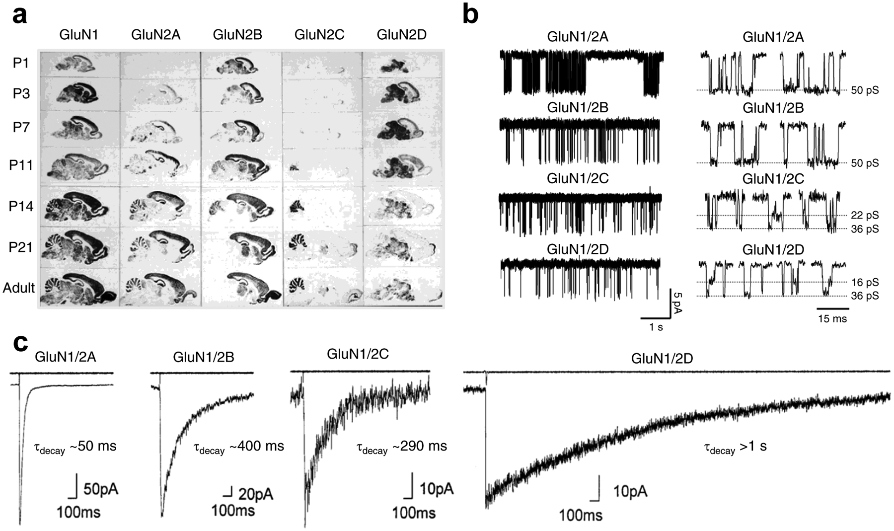
Figure 2. GluN2 subunit-specific expression and functional properties of recombinant NMDA receptor subtypes.

a) Regional and developmental expression of GluN2 subunits in rat brain revealed in autoradiograms using in situ hybridizations of oligonucleotide probes for the relevant mRNAs to parasagittal sections. Modified with permission from Akazawa et al. [92]. b) Single-channel recordings of currents from diheteromeric NMDA receptor subtypes expressed in HEK293 cells (outside-out membrane patches). Open probability is ~0.5 for GluN1/2A, ~0.1 for GluN1/2B, and <0.05 for GluN1/2C and GluN1/2D. Highlights of individual openings are shown on the left. GluN1/2A and GluN1/2B have higher channel conductance (~50 pS) compared to GluN1/2C (~22 and ~36 pS) and GluN1/2D (~16 and ~36 pS). Adapted with permission from Yuan et al. [524]. c) Whole-cell patch-clamp recordings of responses from brief application of glutamate (1 ms of 1 mM glutamate) to recombinant diheteromeric NMDA receptor subtypes expressed in HEK293 cells. The open tip current indicating the duration of the drug application is shown in the upper trace. Adapted with permission from Vicini et al. [62].
