
Fig.4. Therapeutic benefit of combining oHSV with γ-secretase inhibitors.
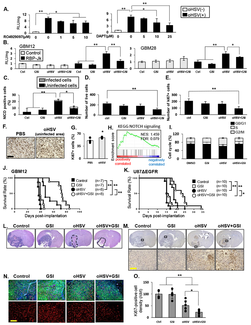
A: NOTCH reporter activity in U87EGFR NOTCH reporter cells after infection with 34.5 ENVE virus (MOI = 0.1) and treated with increasing dose of GSIs; RO4929097 or DAPT for 12 hours. Data shown are mean RLU ± SD (n≥ 3). B: Indicated glioma cells were treated with RO4929097 (10µM), and/or 34.5ENVE (MOI=0.1), and then overlaid with LN229 NOTCH reporter cells. Mean luciferase activity relative to no treatment ± SD is shown (n≥ 3). C: Quantification of GBM28 cells, 9 hours after infection with 34.5ENVE for number of cells staining positive for NICD with or without GSI. Data shown are the mean ± SD. D-E: GBM28 were treated with or without virus in the presence or absence of GSI and incubated for four days. Mean number of viable and proliferating (ki67-positive) cells ± SD (n=4) was measured using flow cytometry. F-G: Immunohistochemistry of ki67 in tissue from U87ΔEGFR subcutaneous mice model treated with PBS (n=4) or oHSV (n=5). Quantification of ki67 density in tumor adjacent to oncolytic virus destroyed plaques were compared to Ki67 density in random sections in untreated tumors. H: GSEA for KEGG_NOTCH signaling by ki67 expression in TCGA GBM database (n=416). I: GSI attenuated oHSV-mediated cell cycle progression. LN229 were treated as indicated for 72 hours. Cells in G0/G1, S and G2/M phases were determined by flow cytometry. Data shown are the mean ± SD (n=3). J-K: Kaplan-Meier analysis of mice implanted with orthotopic GBM12 (J) or U87ΔEGFR (K) cells treated with oHSV with or without GSI therapy. L: Representative hematoxylin and eosin staining of mice bearing GBM12 tumors treated as above, sacrificed twenty days after virus treatment. Both oHSV monotherapy and combination therapy group showed large necrotic areas (dashed line). M: Immunohistochemistry of GBM12 xenograft model showed HSV-1 replication in both of oHSV alone and combination treated group. N-O: Representative fluorescent microscopy images of GBM12 xenograft model immuno-stained for ki67 (red) and human HLA (green) and DAPI (blue) shows nuclear staining. Quantification of ki67 staining overall in entire tumor sections from mice treated as indicated. Data shown are average number of Ki67 +ve cells/ view field in randomly selected view fields/section. Immuno-fluorescent staining of GBM12 xenograft model showed a significant decrease in ki67-positive cell density in combination treated group. Data are shown as the mean ± SD (n = 4-6 in each group). *P<0.05, **P<0.01. Scale bar, 100µm