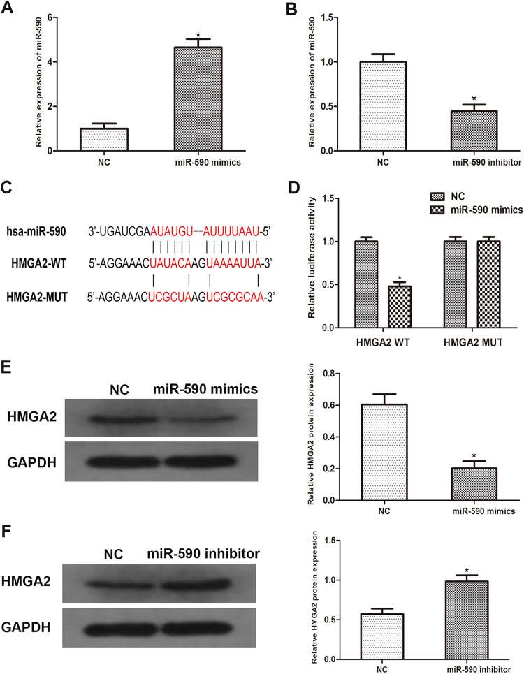
Figure 2.
MiR-590 directly binds to HMGA2 in PDAC samples. A and B, PCR results showed miR-590 mimics or inhibitors were transfected into Capan-2 cells successfully. C, Sequence alignment of predicted miR-590 binding sites with the HMGA2 3′UTR and the mutated sequence of miR-590. D, Luciferase reporter assay was performed in Capan-2 cells that were co-transfected with miR-590 mimics and reporter vectors that containing HMGA2 3′UTR or mutated HMGA2 3′UTR. Relative luciferase activities are shown. E and F, Western blot analysis reveals that the protein level of HMGA2 increased significantly in miR-590 overexpressed cells than miR-590 knockdown cells. Data are presented as means ± SD of 3 independent experiments (*P < .05 compared to control). HMGA2 indicates High mobility group AT-hook 2; miR, microRNA; PDAC, pancreatic ductal adenocarcinoma; PCR, polymerase chain reaction; 3′UTR, 3′untranslated region.

