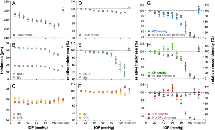
Figure 5.
Effect of IOP elevation on retinal thickness and their relationship to vessel area. (A) Average (±SEM, n = 14) total retinal thickness plotted as function of IOP. (B) Average RNFL IPL thickness versus IOP. (C) Average INL and OPL thickness versus IOP. (D) Total retinal thickness relative to baseline (%) plotted as function of IOP. (E) Relative (%) RNFL and IPL thickness versus IOP. (F) Relative (%) INL and OPL thickness versus IOP. (G) Relative SVC vessel density overlaid with relative thickness of combined RNFL, GCL, and IPL. (H) Relative ICP vessel density over and laid with relative thickness of combined IPL and INL. (I) Relative deep capillary plexus (DCP) vessel density overlaid with relative thickness of combined INL and OPL.

