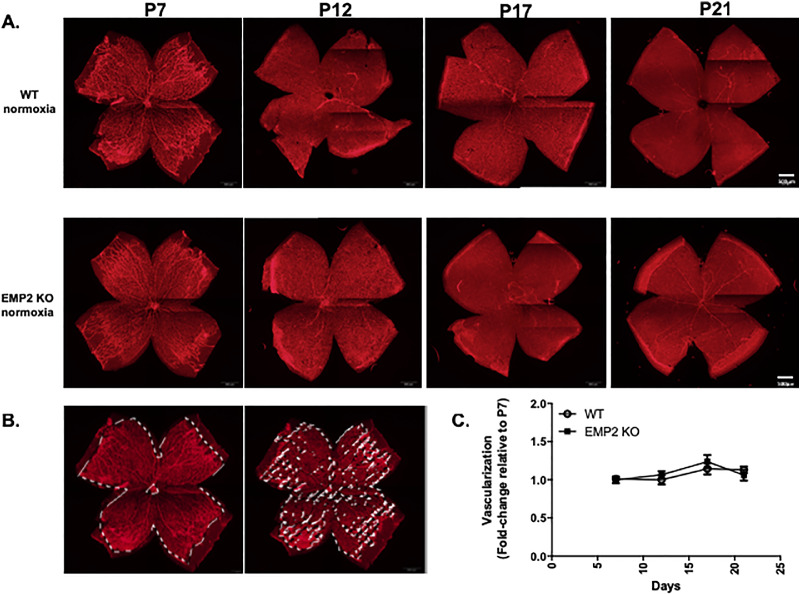
Figure 2.
EMP2 KO does not affect retinal vasculature under physiologic conditions. (A) Representative whole mount images of retinal vasculature in WT (top row) and Emp2 KO (bottom row) mice at P7, P12, P17, and P21. Red staining for lectin outlines endothelial cells. Scale bars represent 500 µm. (B) Descriptive images outlining areas where pixels highlighting retinal vascularization were quantitated. (C) Quantitation of retinal vasculature in whole mount WT and Emp2 KO mice at P7, P12, P17, and P21. Change in retinal vasculature is calculated at P12, P17, and P21 as fold change of vasculature at P7. Data are presented as the mean ± SEM. No differences were observed between the two groups over time (P = 0.6; 2-way ANOVA).

