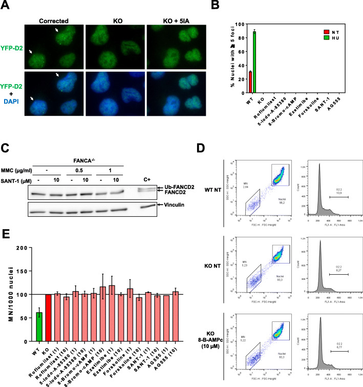
Fig. 3.
Hit validation. a FANCD2 foci detection from YFP-FANCD2 fluorescence from corrected (left lanes), FANCA-deficient (KO, middle lanes), and FANCA-deficient U2OS cell line, incubated with one of the hits (right lanes). Cells were treated with selected hits at 10 μM, 4–6 h later treated with 2 mM HU, and left 16–24 h. Cells were then fixed and immunostained with FANCD2 antibody (see materials and methods). Upper images show FANCD2 localization and bottom images show overlay of FANCD2 with DAPI. White arrows on left lanes show cells with FANCD2 foci. b Graph quantification of cells with 5 or more foci, from the experiment performed in a (and data not shown). Results show mean percentage of cells with foci +/− SEM from three independent experiments with similar results. c FANCA-deficient U2OS cells were treated with SANT-1 at 10 μM, and or MMC, for 24 h. Cells were lyzed and FANCD2 monoubiqutination analyzed by Western blot. Vinculin was used as a loading control. Similar results were obtained from the other hits testes (data not shown). d Spontaneous chromosomal fragility analysis in FANCA-deficient lymphoblastoid cell lines. Graphs show gates from cell cytometry experiments for micronuclei production (left) and G/M cell cycle arrest (right) in Wild type (WT, upper), FANCA-deficient (KO, middle) or FANCA-deficient lymphoblastoid cells stimulated with 10 μM 8-Bromo-AMPc (bottom). e Graph showing mean +/− SEM spontaneous micronuclei production after stimulation of selected hits (at 1 or 10 μM), from at least three independent experiments as in d

