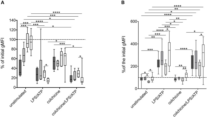
Figure 5.
Alteration of surface markers after stimulation of neutrophils derived from MEFV mutation carriers and patients with other inflammatory diseases. Isolated neutrophils from FMF patients (n = 10, dark gray), heterozygous mutation carriers (n = 6, middle dark gray), controls (n = 7, light dark gray), and patients with other inflammatory diseases (n = 11, white) were cultured as described in Figure 2. After 5 h, CD62L (A) and CD11b (B) expressions were analyzed. Box-and-whisker plots depict 5th−95th percentiles. Significance was analyzed by Kruskal–Wallis followed by Dunn's multi-comparison test, *p < 0.05, **p < 0.01, ***p < 0.001, ****p < 0.0001.

