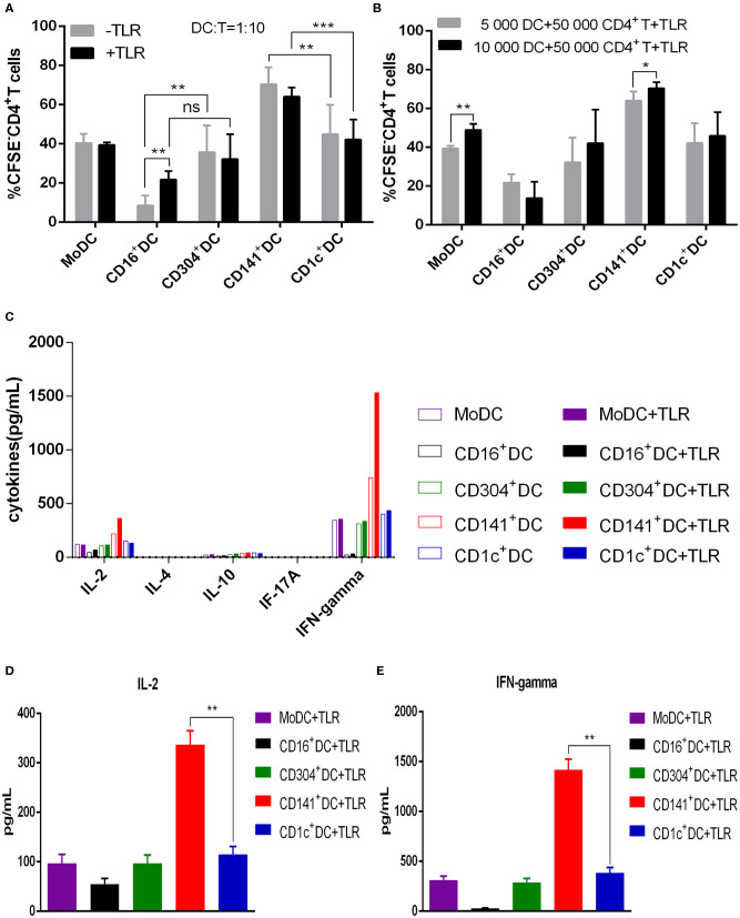
Figure 3.
BDCA3+ (CD141+) DCs have a superior capacity to induce the proliferation of allogeneic CD4+ T cells. (A) Different DC subsets were cultured with CFSE-labeled allogeneic naïve CD4+ T cells in the presence or absence of TLR agonists at a 1:10 for 6 days. Then, T cell proliferation was assessed by flow cytometry. Results of five independent experiments are shown. (B) A total of 5,000 or 10,000 dendritic cells from different DC subsets were cultured with CFSE-labeled allogeneic naïve CD4+ T cells at a 1:10 or 1:5 ratio in the presence of TLR agonists. T cell proliferation was assessed by flow cytometry after 6 days. Results of six independent experiments are shown. (C) Dendritic cells from different DC subsets were cultured for 6 days with allogeneic naïve CD4+ T cells in the presence or absence of TLR agonists at a 1:10 ratio. Cytokine secretion was measured in the supernatant. One experiment out of 3 is shown. (D,E) Secretion of IL-2 (D) and IFN-γ (E) in the allogeneic MLR cultures after 6 days. Results of three independent experiments are shown. DCs were stimulated with or without the following TLR ligands: 3 μg/mL R848 for CD1c+ DCs, 10 μg/mL Poly I:C for BDCA3+ (CD141+) DCs, 1 μg/mL LPS for CD16+ DCs and MoDCs, and 10 μg/mL CpG for pDCs. *P < 0.05, **P < 0.01, and ***P < 0.001. NS, no significance, meaning p > 0.05.

