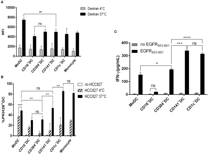
Figure 4.
Capacity of antigen uptake and soluble antigen presentation by human DC subsets. (A) CD1c+ DCs, BDCA3+ (CD141+) DCs, CD16+ DCs, CD304+ DCs, and MoDCs were cultured for 1 h in the presence of 100 μg/mL FITC-dextran at 4 and 37°C, and then the mean fluorescence (MFI) of FITC+DCs was analyzed by flow cytometry. Results of three independent experiments. (B) Uptake of PKH-26-labeled HCC827 (the PKH26 labeled tumor cells were freeze-thawed ×3) by CD1c+ DCs, BDCA3+ (CD141+) DCs, CD16+ DCs, CD304+ DCs, MoDCs, and monocytes after 12 h at 4 and 37°C by flow cytometry. Results of three independent experiments. (C) Five DC subsets from HLA-A*0201+ healthy donors were loaded with HLA-A*0201-restrictive EGFR853−861 peptide for 2 h and used to stimulate an EGFR853−861-specific CTL line for 24 h. Then, the specific IFN-γ production was measured in the supernatant. Results of three independent experiments. *P < 0.05, **P < 0.01, ***P < 0.001, and ****P < 0.0001. NS, no significance, meaning p > 0.05.

