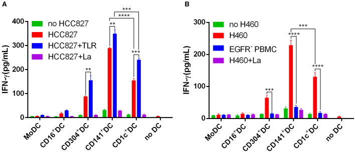
Figure 6.
BDCA3+ (CD141+) DCs strongly and effectively cross-present antigens (EGFR853−861 peptide) from necrotic lung cancer cells. (A) Equivalent numbers of CD1c+ DCs, BDCA3+ (CD141+) DCs, CD16+ DCs, CD304+ DCs, and MoDCs from HLA-A*0201 healthy volunteers were incubated for 18 h with necrotic HCC827 cells at a 1:1 ratio and then used to stimulate an EGFR853−861-specific CD8+ T cell line at a 1:10 ratio in the presence or absence of TLR agonists [3 μg/mL R848 for CD1c+ DCs, 10 μg/mL Poly I:C for BDCA3+ (CD141+) DCs, 1 μg/mL LPS for CD16+ DCs and MoDCs, and 10 μg/mL CpG for pDCs]. Cross-presentation was measured as IFN-γ production by the T cell line. Cross-presentation is proteasome dependent, as it is inhibited by lactacystin (HCC827+La). Results of three independent experiments are shown. (B) Equivalent numbers of CD1c+ DCs, BDCA3+ (CD141+) DCs, CD16+ DCs, CD304+ DCs, and MoDCs from HLA-A*0201 healthy volunteers were incubated for 18 h with necrotic H460 cells at a 1:1 ratio and then used to stimulate an EGFR853−861-specific CD8+ T cell line at a 1:10 ratio in the presence or absence of TLR agonists [3 μg/mL R848 for CD1c+ DCs, 10 μg/mL Poly I:C for BDCA3+ (CD141+) DCs, 1 μg/mL LPS for CD16+ DCs and MoDCs, and 10 μg/mL CpG for pDCs]. Cross-presentation was measured as IFN-γ production by the T cell line. Cross-presentation is proteasome dependent, as it is inhibited by lactacystin (H460+La). EGFR−PBMCs were used as negative control. The results from 3 experiments are shown. *P < 0.05, **P < 0.01, ***P < 0.001, and ****P < 0.0001.

