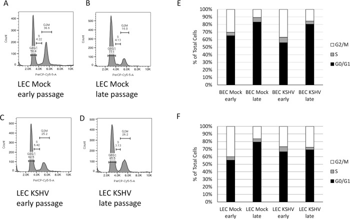
Fig 3. Senescent cells arrest in G0/G1.
Early and late passage mock and KSHV-infected BECs and LECs were harvested, fixed, and stained with propidium iodide. Cell cycle was analyzed by flow cytometry. A-D) Representative histograms from early passage (A and C) and late passage (B and D) mock (A and B) and KSHV-infected (C and D) LECs showing percentage of cells in each phase of the cell cycle. E) Quantification of the percentage of cells in each phase of the cell cycle of early and late passage mock- and KSHV-infected BECs from a representative experiment. F) Quantification of the percentage of cells in each phase of the cell cycle of early and late passage mock- and KSHV-infected LECs from a representative experiment. This experiment was performed three times with similar results.

