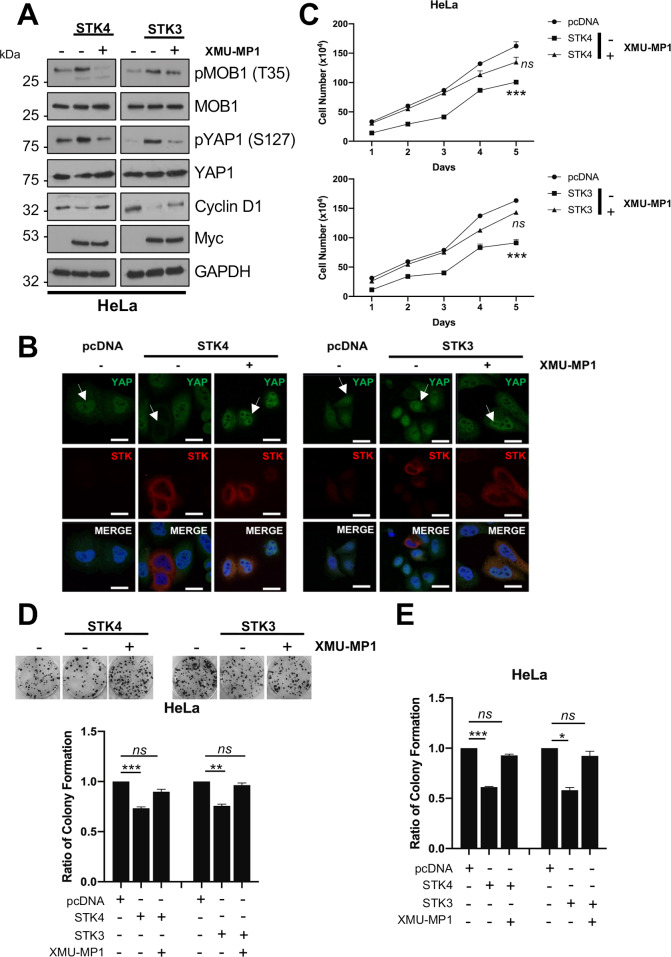
Fig 3. Inhibition of STK4/3 kinase activity prevents the block on proliferation and tumourigenesis.
A) Representative western blots of STK4/3 overexpression in HeLa cells with or without treatment with XMU-MP1 for 8 hours prior to lysis. Lysates were analysed for the phosphorylation of the STK4/3 substrate MOB1, the downstream target YAP and the YAP target gene cyclin D1. The Myc epitope was used to detect fusion protein expression. GAPDH was used as a loading control. B) Immunofluorescence analysis of STK4/3 overexpression in HeLa cells with or without treatment with XMU-MP1 for 8 hours prior to analysis. Cover slips were stained for STK4/3 (red) and YAP1 (green). Nuclei were visualised using DAPI (blue). Images were acquired using identical exposure times. Scale bar, 20 μm. C) Growth curve analysis of HeLa cells overexpressing STK4/3 with or without treatment with XMU-MP1 for 8 hours prior to re-seeding (n = 3). D) Colony formation assay (anchorage dependent growth) of HeLa cells overexpressing STK4/3 with or without treatment with XMU-MP1 for 8 hours prior to re-seeding (n = 3). E) Soft agar assay (anchorage independent growth) of HeLa cells overexpressing STK4/3 with or without treatment with XMU-MP1 for 8 hours prior to re-seeding (n = 3). Error bars represent the mean +/- standard deviation of a minimum of three biological repeats. *P<0.05, **P<0.01, ***P<0.001 (Student’s t-test).

