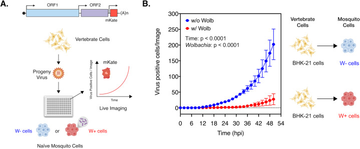
Fig 1. Presence of Wolbachia reduces spread of vertebrate-derived viruses in naïve mosquito cells.
(A) Schematic representation of the experiment. CHIKV expressing mKate fluorescent protein from a second sub-genomic promoter was grown in BHK-21 cells. These progeny viruses were then used to infect naïve C710 cells with (depicted in red) and without (depicted in blue) Wolbachia (wStri strain) synchronously at an MOI of 1 particle/cell. Virus growth in cells, plated on a ninety-six-well plate, was measured in real time by imaging and quantifying the number of red cells (Virus Positive Cells/Image) expressing the virus encoded mKate protein over a period of 48 hours, using live cell imaging. (B) Color of the data points distinguish the two destination cell lines where virus replication was assayed on; blue represent C710 cells without Wolbachia while red represent C710 cells with Wolbachia. Y-axis label (Virus Positive Cells/Image) represent red cells expressing virus-encoded mKate fluorescent protein in a single field of view, four of which were averaged/sample at every two-hour time point collected over the course of infection. Two-way ANOVA with Sidak’s post-hoc test. Error bars represent standard error of mean (SEM) of biological replicates (n = 7–9).

