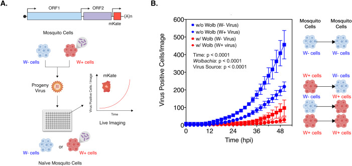
Fig 4. Progeny viruses derived from Wolbachia colonized cells replicate poorly in naïve mosquito cells.
(A) Schematic representation of the experiment. CHIKV expressing mKate fluorescent protein from a second sub-genomic promoter was grown in C710 Aedes albopictus cells in the presence (W+ virus) or absence (W- virus) of Wolbachia (wStri strain). These progeny viruses were then used to infect naïve C710 cells with (depicted in red) and without (depicted in blue) Wolbachia (wStri strain). synchronously at an MOI of 5 particles/cell. Virus growth in cells, plated on a ninety-six-well plate, was measured in real time by imaging and quantifying the number of red cells (Virus Positive Cells/Image) expressing the virus encoded mKate protein over a period of 48 hours, using live cell imaging. (B) Color of the data points distinguish the two destination cell lines where virus replication was assayed on; blue represent C710 cells without Wolbachia while red represent C710 cells with Wolbachia. Shape of the data points refer to the nature of the progeny viruses used to initiate infection; squares represent W- viruses, grown in C710 cells without Wolbachia, while circles represent W+ viruses, grown in C710 cells with Wolbachia. Y-axis label (Virus Positive Cells/Image) represent red cells expressing virus-encoded mKate fluorescent protein in a single field of view, four of which were averaged/sample at every two-hour time point collected over the course of infection. Three-way ANOVA with Tukey’s post-hoc test. Error bars represent standard error of mean (SEM) of biological replicates (n = 9).

