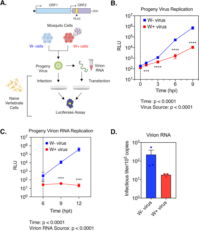
Fig 6. RNA encapsidated within progeny viruses derived from Wolbachia colonized cells are less infectious.
(A) Schematic of experiments performed using Sindbis Virus (SINV) carrying a translationally fused nanoluciferase (nLuc) gene in the second open reading frame (ORF2). (B) Sindbis nLuc reporter viruses (SINV-nLuc) derived from RML12 mosquito cells with (W+ virus) or without (W- virus) Wolbachia (wMel strain) were subsequently used to synchronously infect naïve BHK-21 cells at equivalent MOIs of 5 particles/cell (n = 6). Cell lysates were collected at indicated times post infection and luciferase activity (RLU) was measured and used as a proxy to quantify viral replication. Two-way ANOVA with Tukey’s post-hoc test. Vertical dashed line indicates the first time point collected following removal of the initial inoculum. (C) Replication kinetics of virion encapsidated RNA isolated from progeny SINV-nLuc viruses derived from RML12 mosquito cells with (W+ virus) or without (W- virus) Wolbachia (wMel strain) was determined by measuring luciferase activity (RLU) following transfection of 105 copies of virion encapsulated RNA into BHK-21 cells (n = 6). Two-way ANOVA with Tukey’s post-hoc test. Vertical dashed line at 6 hours post transfection (hpt) indicates the first time point collected following removal of the initial transfection mixture. (D) Infectious titer generated from the aforementioned virion RNAs was determined by counting the number of plaques produced after 48 hours post transfection into BHK-21 cells. Error bars represent standard error of mean (SEM) of biological replicates (n = 3). **P < 0.01, ***P < 0.001, ****P < 0.0001.

