Figure 3.
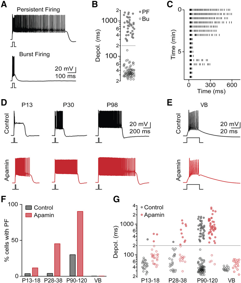
PF occurs late in development and regulated by SK conductances. A, Brief depolarizing current steps (25 ms, 400 pA, RMP = −75 mV) lead to two distinct firing patterns: PF (top) or burst firing (Bu, bottom). B, Summary data (n = 111 neurons) showing bimodal distribution of current-step-evoked membrane depolarization durations. Gray line indicates criterion separating PF and Bu. C, Raster plot of AP activity in representative TRN neuron showing rundown of PF. D, Representative recordings of TRN neurons with PF at three developmental stages, in control (black) and in the presence of apamin (500 nm, red). E, Representative recordings of VB cells, in control (black) and in the presence of apamin (red) show lack of PF in both conditions. F, Summary data showing incidence of PF in TRN across development (P13–P18: n = 24 in control, n = 25 in apamin; P28-P38: n = 23 in control, n = 22 in apamin; P90–P120: n = 111 in control, n = 50 in apamin) and in VB (n = 15 in control, n = 16 in apamin). G, For the same cells as in F, summary data quantifying membrane depolarization duration across development, in control conditions and in apamin.

