Figure 2.
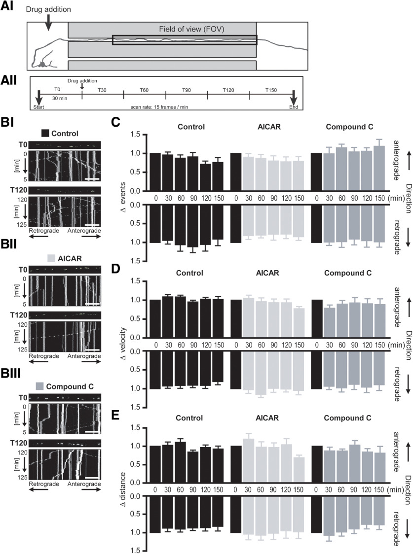
AMPK activation at the somatodendritic region did not alter mitochondrial mobility within the spatially isolated axon segment. A, Schematic of experimental design and time course. AI, Drug addition occurred at the somatodendritic compartment (indicated by the arrow) while mitochondrial mobility was measured in the spatially isolated distal axon segment contained within the microgroove of the microfluidic device (FOV). AII, Mito-GFP fluorescence in this axon segment was imaged at a rate of 15 frames/min (0.25 Hz) for 3 h, with drug addition to the somatodendritic compartment after 30 min (T0) baseline recording. B, Representative kymographs of mitochondrial mobility in the axon segment before (T0) and 2 h (T120) after treatment at the somatodendritic compartment with (BI) experimental buffer (Control), (BII) 0.1 mm AICAR, or (BIII) 10 μm CC. Mito-GFP-tagged mitochondria can be visualized in the still image shown above each kymograph (first image of corresponding time interval). Scale bars, 20 μm. C, The frequency of both anterograde and retrograde mitochondrial moving events within the axon segment remained unaltered by AMPK activation (AICAR) or inhibition (CC) at the somatodendritic region. Mean velocity (D) and distance (E) of mitochondrial movements in both anterograde and retrograde directions remained relatively constant within the spatially isolated distal axon segment during AICAR or CC treatment at the somatodendritic region. Repeated-measures ANOVA, Dunnett's multiple-comparison post-test. n ≥ 6 experiments from at least four independent culture preparations.

