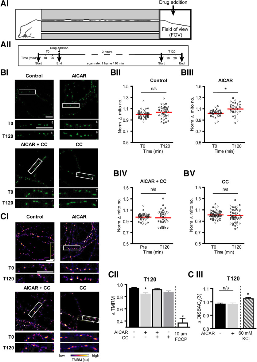
Figure 4.
Localized AMPK activation at the distal axon induced an accumulation of mitochondria directly at the site of treatment. A, Schematic of experiment design. Fluorescent confocal images were captured at the distal axon (FOV) for 30 min at a rate of 1 frame/10 min, 30 min before (T0) and 2 h after (T120) treatment with 0.1 mm AICAR ± 10 μm CC directly to this region. BI, Representative images (354.2 µm × 354.2 µm) of mito-GFP-positive mitochondria within the axonal compartment directly exposed to treatment. Section of distal axon (white box) expanded below to show the number of mitochondria before (T0) and 2 h after treatment (T120). Mitochondrial counts for the section noted in the top right-hand corner of each image. Scale bars, 20 µm. BII-BV, Quantification of the mean number of mitochondria in the distal axon before and 2 h after AICAR ± CC treatment at this region. AICAR treatment (BIII) at the distal axon resulted in an accumulation of mitochondria in this region after 2 h, an effect removed by cotreatment with CC (BIV). *p ≤ 0.001 (two-tailed paired t test). n ≥ 32 FOVs from at least four independent culture preparations. C, Cortical neurons were preloaded with 20 nm TMRM for 30 min before imaging. CI, Representative images of mitochondrial TMRM fluorescence levels within the axonal compartment at the start of the experiment. Section of the distal axons within the white box is expanded below to show TMRM fluorescence before (T0) and 2 h after treatment (T120). Scale bars, 20 µm. CII, AICAR treatment resulted in a mild decrease in mitochondrial membrane potential, compared with control. FCCP, a potent uncoupler of oxidative phosphorylation, was used as a positive control. *p ≤ 0.05 compared with control levels at T150 (one-way ANOVA with Dunnett's multiple-comparison post hoc test). n ≥ 32 FOVs from at least four independent culture preparations. FCCP data were obtained from 23 FOVs from two experiments. CIII, Cells were preloaded with 1 μm DiSBAC4(3) for 30 min before imaging, to visualize changes in the plasma membrane potential. The 2 h AICAR treatment did not alter the plasma membrane potential. Two-tailed unpaired t test. n = 69 FOVs from at least four independent culture preparations. Potassium chloride (KCl, 60 mm) was used as a positive control for depolarization of the plasma membrane, resulting in a significant increase in DiSBAC4(3) fluorescence. KCl data were obtained from 40 FOVs from four experiments. *p ≤ 0.05 compared with control levels at T150.

