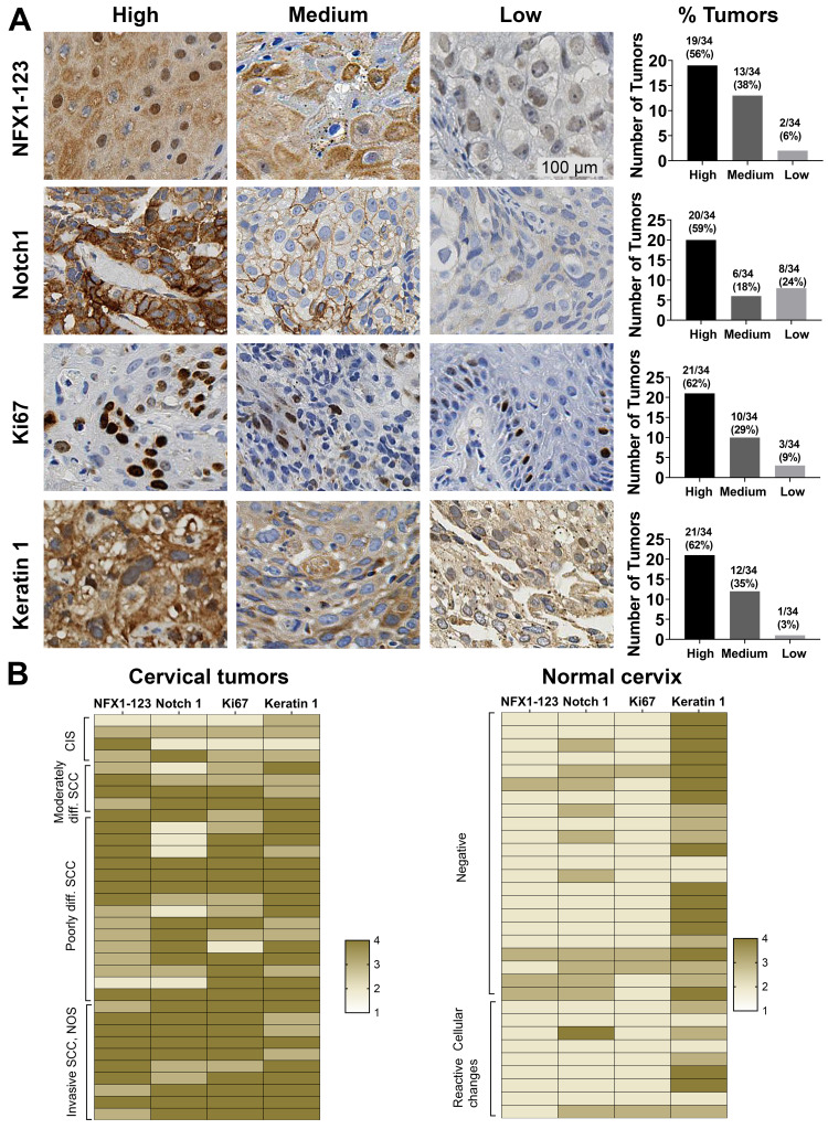
Figure 1.
NFX1-123 and proliferation and differentiation marker protein expression in HPV16 positive cervical cancer tissues. (A) Immunohistochemical staining of NFX1-123, Notch1, Ki67, and Keratin 1 in HPV16 positive cervical tumors (n = 34). Representative images of high, medium, and low staining are shown for each protein. Number of tumors and corresponding percent of total tumors graded as high, medium, or low staining were quantified and shown in graphs (right). (B) Heat map representation of the NFX1-123, Notch1, Ki67 and Keratin 1 expression in cervical cancer samples (n = 34) and normal cervical epithelial samples (n = 31). The cervical tumors are clustered by histologic classification (carcinoma in situ (CIS), moderately differentiated, poorly differentiated, or invasive squamous cell carcinomas (SCC)) and the normal cervical tissues clustered by cytologic classification (negative or reactive cellular changes). The staining intensity of both the HPV 16 positive cervical cancers and the normal cervical specimens were converted to a four-point colorimetric scale: 1= non-detected, 2 = low, 3 = medium, 4 = high.

